The CIC nanoGUNE develops a technique to differentiate the protein structure more in detail than ever
2013/12/18 Carton Virto, Eider - Elhuyar Zientzia Iturria: Elhuyar aldizkaria
The structure of the protein is fundamental for the protein to fulfill its function. Likewise, the secondary space structure that adopts protein (alpha helix or beta page) plays an important role in some neurodegenerative diseases. Consequently, the development of detection techniques at such a detailed scale of these secondary structures can be of great help in research.
The method, developed by a team of researchers from CIC nanoGUNE, the Free University of Berlin and the Nefere Institute, has published its results in the journal Nature Communications.
Maps at nanometric scale
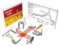
The technique developed is based on infrared nanospectroscopy (nano-FTIR). This is an optical technique that combines the near field scanning optical microscope (s-SNOM) with infrared spectroscopy (FTIR) by Fourier transformation. The nano-FTIR spectroscopy illuminates a metal tip pointing to a broadband infrared laser and analyzes the retrospective light with a specially designed transformed Fourier spectroscope.
_galeria.jpg)
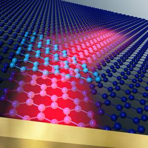
Being a common tool to study the secondary structure of proteins, it does not allow to make a map on a nanometric scale of proteins. The researchers of CIC nanoGUNE have refined this technique obtaining a resolution less than 30 nm. “The tip is a kind of antenna for infrared light that wraps the light in the tip. The nanofoco of this upper peak can be considered as an ultratxiki source of infrared light. It is so small that it only illuminates an area of 30x30 nm, which is the scale of large protein complexes,” explains Rainer Hillenbrand.
The researchers have studied with the new technique individual viruses, ferritin complexes and infrared spectra of small insulin fibers, distinguishing structures that would not reach standard spectroscopy. “In a mixture of insulin fibers and viruses, standard FTIR spectroscopy did not detect the presence of alpha-heliz virus,” said group biologist Simon Poly. They have also been able to measure the infrared spectrum of a single ferritin particle: They are complex of 24 very low mass proteins, only 1 attograms (10-18 g); the researchers have been able to clearly differentiate the alpha helix structures.
According to the researchers, one aspect of great practical importance is that the nano-FTIR spectrum combines very well with the traditional FTIR spectrum and that spatial resolution increases more than 100 times conventional infrared spectroscopy. Hillenbrand has described as "exciting" the new possibilities offered by the nano-FTIR technique. “With the sharpest tips and the improvement of the function of antennas we hope that in the future the infrared spectrum of unique proteins will be obtained,” he said.

Gai honi buruzko eduki gehiago
Elhuyarrek garatutako teknologia


